
File:Anatomy of Human Ear with Cochlear Frequency Mapping.svg

Size of this PNG preview of this SVG file: 674 × 519 pixels. Other resolutions: 312 × 240 pixels | 624 × 480 pixels | 998 × 768 pixels | 1,280 × 986 pixels | 2,560 × 1,971 pixels.
Original file (SVG file, nominally 674 × 519 pixels, file size: 33 KB)
| This is a file from the Wikimedia Commons. Information from its description page there is shown below. Commons is a freely licensed media file repository. You can help. |
Summary
| Description |
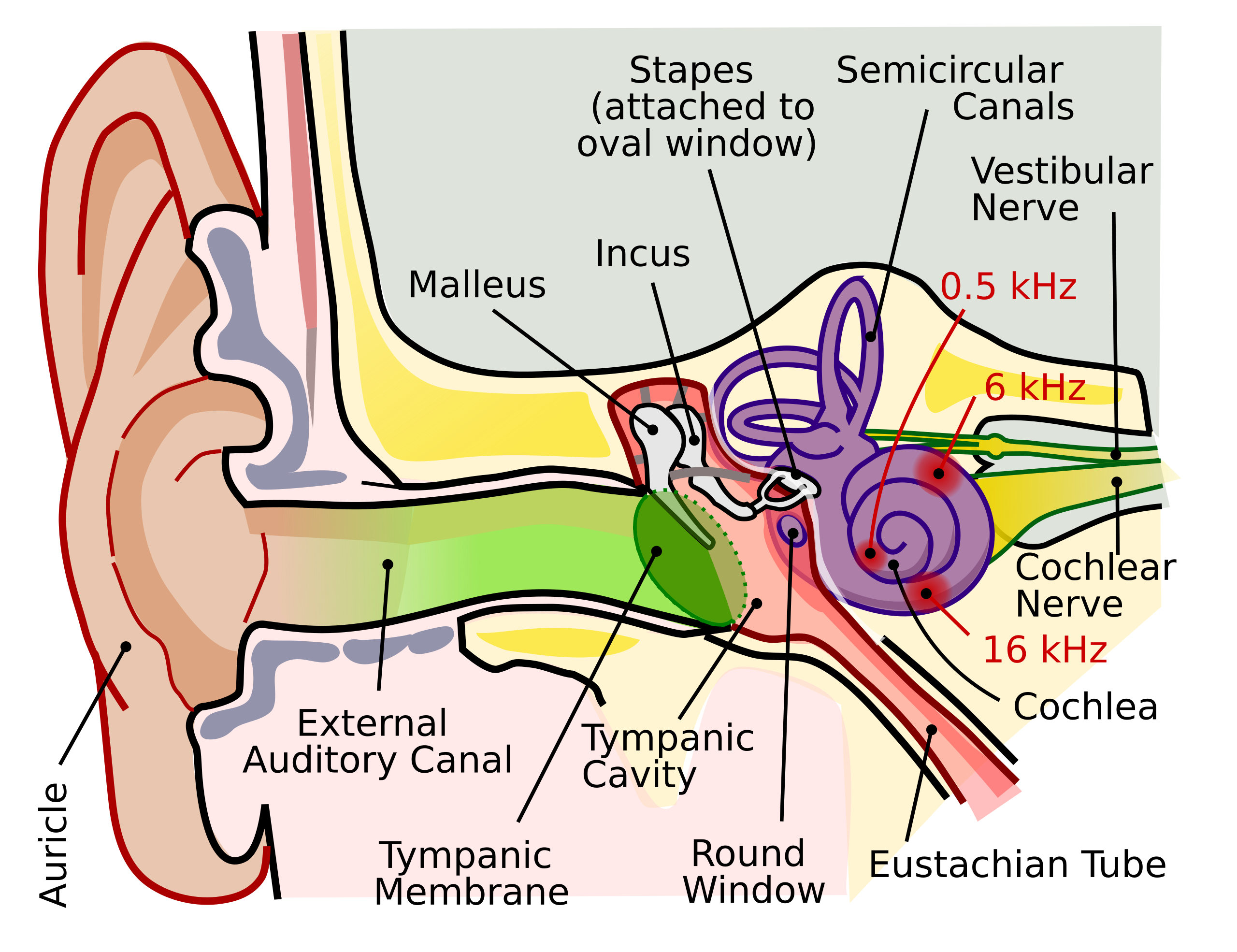
English: The human ear and frequency mapping in the cochlea. The three ossicles incus, malleus, and stapes transmit airborne vibration from the tympanic membrane to the oval window at the base of the cochlea. Because of the mechanical properties of the basilar membrane within the snail-shaped cochlea, high frequencies will produce a vibration peak near the oval window, whereas low frequencies will stimulate receptors near the apex of the cochlea (locations for three frequencies indicated schematically). Information from the cochlear receptor cells is transmitted to the cochlear nuclei via the 8th cranial nerve, and on through the midbrain to the cortex. |
| Date | |
| Source | Own work (Original text: Own work by uploader, derived from File:Anatomy_of_the_Human_Ear.svg) |
| Author | Inductiveload |
| Permission (Reusing this file) |
This file is licensed under the Creative Commons Attribution-Share Alike 2.5 Generic license.
|
| Other versions |
[]
|
| SVG development | This vector image was created with Inkscape. This file is translated using SVG switch elements: all translations are stored in the same file. |


File history
Click on a date/time to view the file as it appeared at that time.
| Date/Time | Thumbnail | Dimensions | User | Comment | |
|---|---|---|---|---|---|
| current | 22:29, 16 September 2018 | | 674 × 519 (33 KB) | wikimediacommons>JoKalliauer | added systemLanguage="eo" |
File usage
The following page uses this file: