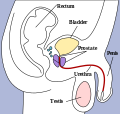
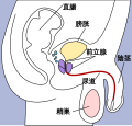
File:Male pelvic structures.svg

Size of this PNG preview of this SVG file: 270 × 257 pixels. Other resolutions: 252 × 240 pixels | 504 × 480 pixels | 807 × 768 pixels | 1,076 × 1,024 pixels | 2,152 × 2,048 pixels.
Original file (SVG file, nominally 270 × 257 pixels, file size: 12 KB)
File history
Click on a date/time to view the file as it appeared at that time.
| Date/Time | Thumbnail | Dimensions | User | Comment | |
|---|---|---|---|---|---|
| current | 03:41, 27 March 2007 |  | 270 × 257 (12 KB) | wikimediacommons>Indolences |
File usage
The following 40 pages use this file:
- Acrosome
- Appendix of testis
- Bowman–Heidenhain hypothesis
- Cavernous tissue
- Cortical lobule
- Crossed renal ectopia
- Ductuli transversi
- Ectopic kidney
- Facet cell
- Follicular antrum
- Follicular fluid
- Genitourinary amoebiasis
- Germinal epithelium (male)
- Juxtaglomerular cell
- Lacunae of Morgagni
- Man
- Medulla of ovary
- Medullary ray (anatomy)
- Membrana granulosa
- Mesovarium
- Ovarian fossa
- Parametrium
- Paroophoron
- Pelvic kidney
- Penile pain
- Perineal sponge
- Pre-prostatic urethra
- Prostatic ducts
- Prosthetic testicle
- Renal column
- Renal sinus
- Supernumerary kidney
- Supravaginal portion of cervix
- Theca externa
- Urethral gland
- Urine sodium
- Uterine cavity
- Uterine horns
- Vesicular appendages of epoophoron
- Template:Genitourinary-stub


