File:Spectroscopy overview.svg

Original file (SVG file, nominally 760 × 303 pixels, file size: 2.49 MB)
| This is a file from the Wikimedia Commons. Information from its description page there is shown below. Commons is a freely licensed media file repository. You can help. |
Summary
| DescriptionSpectroscopy overview.svg |
Català: Esquema de l'absorció de radiació electromagnètica.
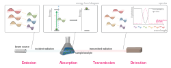
English: An overview of electromagnetic radiation absorption. This example discusses the general principle using visible light as specific example. A white beam source -- emitting light of multiple wavelengths -- is focused on a sample (the complementary color pairs are indicated by the yellow dotted lines. Upon striking the sample, photons that match the energy gap of the molecules present (green light in this example) are absorbed in order to excite the molecule. Other photons transmit unaffected and, if the radiation is in the visible region (400-700nm), the transmitted light appears as its complementary color. By comparing the attenuation of the transmitted light with the incident, an absorption spectrum can be obtained. |
| Date | |
| Source | Own work |
| Author | Jon Chui |
| Other versions |
Derivative works of this file: File:Spectroscopy overview.svg has 3 translations.
Other related versions: [edit]
|
This file is translated using SVG <switch> elements. All translations are stored in the same file! Learn more.
For most Wikipedia projects, you can embed the file normally (without a To translate the text into your language, you can use the SVG Translate tool. Alternatively, you can download the file to your computer, add your translations using whatever software you're familiar with, and re-upload it with the same name. You will find help in Graphics Lab if you're not sure how to do this. |
| This SVG file contains embedded text that can be translated into your language, using any capable SVG editor, text editor or the SVG Translate tool. For more information see: About translating SVG files. |
Licensing
- You are free:
- to share – to copy, distribute and transmit the work
- to remix – to adapt the work
- Under the following conditions:
- attribution – You must give appropriate credit, provide a link to the license, and indicate if changes were made. You may do so in any reasonable manner, but not in any way that suggests the licensor endorses you or your use.
- share alike – If you remix, transform, or build upon the material, you must distribute your contributions under the same or compatible license as the original.

|
Permission is granted to copy, distribute and/or modify this document under the terms of the GNU Free Documentation License, Version 1.2 or any later version published by the Free Software Foundation; with no Invariant Sections, no Front-Cover Texts, and no Back-Cover Texts. A copy of the license is included in the section entitled GNU Free Documentation License.http://www.gnu.org/copyleft/fdl.htmlGFDLGNU Free Documentation Licensetruetrue |
| Annotations InfoField | This image is annotated: View the annotations at Commons |
White light is composed of continuous wavelengths of light in the visible region (400-700nm). Here we simplify its representation with discrete colors arranged in descending order wavelength (i.e., increasing energy). Polarization does not affect absorption and is not represented; complementary colors are denoted with yellow dotted lines.
"Beam source" generally indicates a broad spectrum light, e.g., from black-body radiation. Lasers are not within this scope as they are monochromatic (single wavelength).
Molecules have quantized energy levels, and photons have quantized energy. If the incoming photon has exactly the matching energy for the promotion of the molecule, the molecule would absorb the photon, and change state from the ground state to an excited state. The specific physical change depends on the radiation.
Light in the visible region induces electronic excitation (rearrangement of electron clouds).
This applies only for visible light - the removal of a color results in the beam appearing as its complementary color. Here, the removal of green within the red-green pair gives the transmitted beam a red color.
Note that absorptions always has a non-zero line-width despite the quantized nature of light and matter. This is discussed in the article on line broadening.
A spectrum is effectively a (continuous) histogram describing the composition of light. A spectrum can be represented as a transmission spectrum (shown in gray) where the y-axis shows the photon count of transmitted light, or an absorption spectrum (magenta) where the attenuation is plotted. The former is commonly used in infrared/microwave spectroscopy, and the latter in UV-Vis and NMR.
Historically a detector is an optical device where the beam is split (by a diffraction grating or prism) and captured on a photographic plate. Modern spectroscopy usually uses a CCD array in conjunction with digital electronics.
In the laboratory, this is commonly a sample containing an analyte dissolved in a solvent. Spectroscopy is a general technique - an example on the astronomical scale would involve a star as the "beam source", a planet as the "sample", and a telescope-CCD array as the detector, with interstellar space between each. The concepts and principles remain the same.
Despite the quantized nature of light and matter, absorptions are broad and never of zero line-width. This phenomenon is explained in the article on line broadening.

|
This image has been assessed under the valued image criteria and is considered the most valued image on Commons within the scope: Overview of Absorption Spectroscopy. You can see its nomination here. |
Captions
Items portrayed in this file
depicts
8 January 2011
image/svg+xml
File history
Click on a date/time to view the file as it appeared at that time.
| Date/Time | Thumbnail | Dimensions | User | Comment | |
|---|---|---|---|---|---|
| current | 12:20, 30 September 2023 | 760 × 303 (2.49 MB) | wikimediacommons>Townie | File uploaded using svgtranslate tool (https://svgtranslate.toolforge.org/). Added translation for ca. |
File usage
The following 2 pages use this file:
Metadata
This file contains additional information, probably added from the digital camera or scanner used to create or digitize it.
If the file has been modified from its original state, some details may not fully reflect the modified file.
| Width | 760.243px |
|---|---|
| Height | 302.655px |


